
Introduction
Precision surgery represents a paradigm shift in the field of surgical intervention, emphasizing tailored approaches that consider the unique anatomical and pathological characteristics of each patient. This approach aims to enhance surgical outcomes by minimizing damage to surrounding tissues while maximizing the effectiveness of the procedure. Central to the success of precision surgery is the integration of advanced imaging techniques, which provide critical insights into the patient’s anatomy and pathology.
By leveraging high-resolution images, surgeons can make informed decisions that lead to more effective interventions. Imaging technologies have evolved significantly over the past few decades, transforming the landscape of surgical practice. The ability to visualize internal structures in real time allows for a more nuanced understanding of complex cases, particularly in oncology, neurosurgery, and orthopedic surgery.
As a result, imaging not only aids in diagnosis but also plays a pivotal role in guiding surgical strategies, ultimately leading to improved patient outcomes and reduced recovery times.
Key Takeaways
- Precision surgery relies on advanced imaging techniques for accurate preoperative planning and intraoperative navigation.
- Imaging plays a crucial role in identifying tumor margins and guiding targeted therapies for improved patient outcomes.
- Various imaging techniques such as MRI, CT scans, and PET scans are used to visualize and map out the surgical site in precision surgery.
- Intraoperative navigation using real-time imaging helps surgeons to precisely locate and remove tumors while minimizing damage to surrounding healthy tissue.
- Advancements in imaging technology, such as 3D imaging and augmented reality, are shaping the future of precision surgery by providing more detailed and accurate visualization of the surgical site.
The Importance of Imaging in Preoperative Planning
Preoperative planning is a crucial phase in the surgical process, where detailed assessments are made to determine the best course of action. Imaging serves as a cornerstone in this stage, providing surgeons with a comprehensive view of the surgical site. Techniques such as magnetic resonance imaging (MRI), computed tomography (CT), and ultrasound allow for the identification of tumors, vascular structures, and other critical anatomical features.
This information is essential for developing a tailored surgical plan that addresses the specific needs of the patient. Moreover, imaging facilitates risk assessment by highlighting potential complications that may arise during surgery. For instance, preoperative imaging can reveal the proximity of a tumor to vital structures, enabling surgeons to strategize their approach and minimize risks.
By understanding the spatial relationships between different anatomical components, surgeons can optimize their techniques and improve overall surgical precision. This meticulous planning ultimately contributes to better postoperative outcomes and enhances patient safety.
Imaging Techniques Used in Precision Surgery
A variety of imaging techniques are employed in precision surgery, each offering unique advantages that cater to different surgical needs. Magnetic resonance imaging (MRI) is particularly valuable in soft tissue visualization, making it indispensable in neurosurgery and orthopedic procedures. Its ability to provide detailed images of brain structures or joint tissues allows surgeons to plan their interventions with remarkable accuracy.
Computed tomography (CT) scans are another vital tool, especially in cases involving complex bony structures or when rapid assessment is required. CT imaging provides cross-sectional views that help identify fractures, tumors, or other abnormalities with high precision. Additionally, advancements in 3D imaging technology have enabled surgeons to create three-dimensional models of anatomical structures, further enhancing their understanding of the surgical landscape.
These models can be used for simulation and rehearsal, allowing for a more confident approach during actual procedures.
Role of Imaging in Intraoperative Navigation
| Imaging Modality | Advantages | Challenges |
|---|---|---|
| MRI | High soft tissue contrast, no radiation exposure | Not suitable for intraoperative use, limited availability |
| CT | High spatial resolution, good for bone visualization | Ionizing radiation, limited soft tissue contrast |
| Ultrasound | Real-time imaging, no radiation exposure | Operator-dependent, limited depth penetration |
| Fluoroscopy | Real-time imaging, good for dynamic procedures | Ionizing radiation, limited soft tissue contrast |
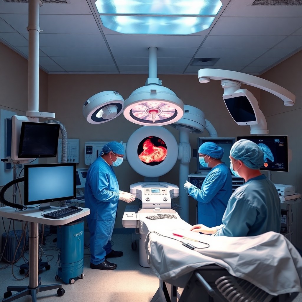
Intraoperative navigation has revolutionized the way surgeries are performed by integrating real-time imaging into the surgical workflow. This technology allows surgeons to visualize their instruments’ position relative to critical anatomical landmarks during the procedure. By utilizing advanced imaging modalities such as fluoroscopy or intraoperative ultrasound, surgeons can make immediate adjustments based on live feedback, significantly enhancing their precision.
The role of imaging in intraoperative navigation is particularly evident in complex surgeries such as spinal operations or tumor resections. For example, during spinal surgery, real-time imaging can guide the placement of screws and rods with unparalleled accuracy, reducing the risk of complications. Similarly, in tumor resections, intraoperative imaging helps ensure complete removal of cancerous tissue while preserving healthy surrounding structures.
This dynamic interaction between imaging and surgical technique exemplifies how technology can enhance precision and improve patient outcomes.
Imaging-Guided Tumor Resection and Targeted Therapies
Imaging-guided tumor resection has become a cornerstone of modern oncology, allowing for more effective removal of malignant tissues while minimizing damage to healthy cells. Techniques such as stereotactic navigation utilize preoperative imaging data to guide surgeons during tumor excision. This method ensures that the tumor is removed with high precision, reducing the likelihood of residual disease and improving long-term survival rates.
In addition to traditional resection techniques, imaging plays a vital role in targeted therapies such as radiofrequency ablation or stereotactic body radiation therapy (SBRT). These approaches rely on precise imaging to deliver treatment directly to the tumor while sparing surrounding healthy tissue. For instance, real-time imaging can help monitor the effectiveness of ablation procedures by visualizing changes in tumor size or structure during treatment.
This integration of imaging into therapeutic strategies exemplifies how technology can enhance both surgical precision and treatment efficacy.
Advancements in Imaging Technology for Precision Surgery
The field of imaging technology has witnessed remarkable advancements that have significantly impacted precision surgery. Innovations such as high-definition endoscopy and intraoperative MRI have enhanced visualization capabilities, allowing surgeons to see finer details than ever before. These technologies enable minimally invasive approaches that reduce recovery times and improve patient comfort while maintaining high levels of precision.
Furthermore, artificial intelligence (AI) is beginning to play a transformative role in imaging analysis. AI algorithms can assist in interpreting complex imaging data, and identifying patterns that may be missed by human eyes. This capability not only enhances diagnostic accuracy but also aids in preoperative planning by predicting potential complications based on historical data.
As these technologies continue to evolve, they promise to further refine the precision of surgical interventions.
Future Directions in Imaging for Precision Surgery
Looking ahead, the future of imaging in precision surgery is poised for exciting developments that will further enhance surgical outcomes. One promising direction is the integration of augmented reality (AR) into surgical practice. By overlaying digital images onto the surgeon’s field of view, AR can provide real-time guidance during procedures, allowing for even greater precision and confidence.
Additionally, advancements in molecular imaging techniques may enable surgeons to visualize not only anatomical structures but also biological processes at a cellular level. This could lead to more personalized surgical approaches tailored to individual patient’s unique tumor biology or anatomical variations. As research continues to push the boundaries of what is possible with imaging technology, the potential for improved precision surgery remains vast and promising.
Conclusion
The role of imaging in enhancing precision surgery cannot be overstated. Advanced imaging techniques are integral to modern surgical practice, from preoperative planning to intraoperative navigation and targeted therapies. As technology continues to evolve, it promises to further refine surgical precision and improve patient outcomes across various medical disciplines.